
Renin Angiotensin System
PATHOPHYSIOLOGICAL REMINDER
1. The Renin-Angiotensin System
The renin-angiotensin system (RAS), also known as the renin-angiotensin-aldosterone system (RAAS), is a major hormonal regulatory mechanism involved in the maintenance of cardiovascular and renal homeostasis. It plays a fundamental role in controlling arterial blood pressure, circulating blood volume, sodium balance, and electrolyte regulation. The system is classically considered a hormonal system involving a coordinated sequence of enzymatic reactions and physiological responses.
The circulating renin, released by the juxtaglomerular cells of the kidneys, acts on angiotensinogen, a precursor protein primarily produced by the liver, to generate angiotensin I. Angiotensin I is subsequently converted into angiotensin II through the action of angiotensin-converting enzyme (ACE), which is predominantly expressed on the surface of endothelial cells, particularly within the pulmonary vascular bed.
Following its formation, angiotensin II is distributed through the bloodstream to different organs and tissues, where it interacts with specific receptors to induce a wide range of physiological responses. These responses include vasoconstriction, stimulation of aldosterone secretion, regulation of renal sodium reabsorption, and modulation of sympathetic nervous system activity.
This circulating pathway, referred to as the extrinsic renin-angiotensin system, is responsible for many of the major systemic physiological effects associated with blood pressure regulation and fluid-electrolyte homeostasis.
However, the renin-angiotensin system is not exclusively restricted to the circulation. Several organs and tissues possess local components of this system, allowing angiotensin peptides to be generated and act independently of the circulating pathway. This tissue-based mechanism is known as the intrinsic renin-angiotensin system or local renin-angiotensin system.
Local renin-angiotensin systems have been identified in the heart, blood vessels, kidneys, brain, and other tissues. They contribute to the regulation of tissue-specific physiological functions, including cardiac contractility, vascular tone, cellular growth, inflammatory responses, extracellular matrix remodeling, and renal function.
The distinction between the extrinsic and intrinsic renin-angiotensin systems is particularly important because local angiotensin II production can contribute to tissue responses even when circulating angiotensin II concentrations remain relatively stable. Consequently, dysregulation of local renin-angiotensin activity may participate in the development of cardiovascular and renal pathological conditions.
2. Angiotensin-Converting Enzyme
Angiotensin-converting enzyme (ACE), also known as ECA (enzyme de conversion de l'angiotensine), is a membrane-associated glycoprotein that plays a central role in the regulation of the renin-angiotensin system. Structurally, ACE comprises two homologous domains, each containing a catalytic site responsible for enzymatic activity.
ACE is mainly present on the membrane surface of endothelial cells, with particularly high expression in the pulmonary vascular bed. Nevertheless, the enzyme is also found in other vascular tissues and can circulate in plasma in a soluble form.
ACE is a non-specific enzyme whose principal physiological substrates include angiotensin I and bradykinin. Its dual enzymatic activity is particularly important because it simultaneously participates in the production of a potent vasoconstrictor and the degradation of a vasodilator peptide.
The conversion of angiotensin I into angiotensin II represents one of the most important reactions catalyzed by ACE. During this process, angiotensin I is converted into the biologically active octapeptide angiotensin II through the removal of two amino acids from its C-terminal end.
It is interesting to note that the discovery of ACE inhibitors in the 1960s was associated with research on peptides derived from the venom of certain vipers. These compounds were found to intensify the biological effects of bradykinin, contributing to the development of the first angiotensin-converting enzyme inhibitors.
Bradykinin is a vasoactive peptide that promotes vasodilation through mechanisms involving the release of nitric oxide and prostacyclin from endothelial cells. By inhibiting its degradation, ACE inhibitors increase bradykinin availability and enhance its vasodilatory effects. This mechanism contributes to their antihypertensive activity.
The conversion of angiotensin I into angiotensin II is extremely rapid under physiological conditions. Consequently, the pharmacodynamic responses associated with these two peptides are difficult to distinguish based solely on their circulating effects. However, the intrinsic biological potency of angiotensin I is considerably lower, representing less than 1% of the potency of angiotensin II.
Alternative Pathways of Angiotensin II Formation
Although ACE is the principal enzyme responsible for converting angiotensin I into angiotensin II in the classical pathway, alternative enzymatic mechanisms also exist within different tissues.
Some tissues contain enzymes capable of converting angiotensinogen into angiotensin peptides through non-renin proteolytic pathways. Other enzymes can directly generate angiotensin II from angiotensinogen or convert angiotensin I into angiotensin II independently of ACE.
Among these enzymes, cathepsin G and tonin have been identified as contributors to alternative angiotensin II-generating pathways. In addition, cathepsin G and chymases can participate in the non-ACE-dependent conversion of angiotensin I into angiotensin II.
Chymases are particularly relevant in cardiovascular and renal physiology. In the heart and kidneys, these enzymes contribute to local angiotensin II formation, providing an alternative mechanism for maintaining tissue renin-angiotensin activity independently of circulating ACE.
The presence of these alternative pathways demonstrates that angiotensin II production is not exclusively dependent on ACE activity. This has important implications for understanding tissue-specific renin-angiotensin regulation and the pharmacological effects of different inhibitors targeting this system.
3. Angiotensin Receptors
The biological effects of angiotensin II are mediated through specific transmembrane receptors belonging to the G protein-coupled receptor (GPCR) family. These receptors are involved in the transmission of extracellular signals into intracellular responses, regulating vascular function, electrolyte balance, cellular proliferation, and cardiovascular remodeling.
Two major angiotensin II receptor subtypes have been identified: AT1 receptors and AT2 receptors. Although both receptors bind angiotensin II, they exhibit limited sequence homology and differ significantly in their intracellular signaling pathways, tissue distribution, and physiological functions.

AT1 Receptors
Most of the classical physiological and pathological effects of angiotensin II are mediated by stimulation of AT1 receptors.
AT1 receptor activation triggers several intracellular signaling pathways, particularly those involving phospholipase C, inositol trisphosphate, diacylglycerol, and intracellular calcium mobilization. These signaling mechanisms contribute to vascular smooth muscle contraction and increased peripheral vascular resistance.
AT1 receptor stimulation also promotes aldosterone secretion from the adrenal cortex, enhances sympathetic nervous system activity, and increases renal sodium reabsorption.
Beyond its immediate hemodynamic effects, AT1 receptor activation is involved in long-term cardiovascular remodeling. Sustained stimulation can promote cellular hypertrophy, fibroblast activation, collagen synthesis, extracellular matrix deposition, and inflammatory signaling.
These effects are particularly important in pathological conditions characterized by chronic activation of the renin-angiotensin system, including hypertension, cardiac hypertrophy, heart failure, and progressive renal injury.
AT2 Receptors
The functional role of AT2 receptors is generally considered different from that of AT1 receptors. These receptors are sometimes described as cardioprotective because several of their biological effects oppose the actions mediated by AT1 receptor stimulation.
AT2 receptor activation is associated with antiproliferative, proapoptotic, vasodilatory, and antihypertensive effects. These properties suggest an important role in regulating tissue growth, vascular function, and cellular responses to angiotensin II.
AT2 receptor signaling can involve pathways that stimulate nitric oxide production, increase cyclic GMP formation, and promote vascular relaxation. Depending on the tissue and experimental conditions, AT2 receptor activation may also influence inflammatory responses, tissue remodeling, and cellular differentiation.
The functional balance between AT1 and AT2 receptor signaling is therefore an important determinant of the overall biological response to angiotensin II.
In physiological and pathological conditions, the relative expression and activation of these two receptor subtypes can influence whether angiotensin II produces predominantly vasoconstrictive and proliferative effects or contributes to vasodilatory and tissue-protective mechanisms.
4. Physiological Effects
The main physiological function of the renin-angiotensin system is to ensure the short-term and long-term regulation of arterial blood pressure. It achieves this through coordinated effects on vascular resistance, renal sodium handling, circulating blood volume, and neurohormonal activity.
Angiotensin II is an extremely potent vasoconstrictor, with an EC50 of approximately 0.3 nM. Its interaction with AT1 receptors on vascular smooth muscle cells produces rapid contraction and an increase in peripheral arterial resistance.
In addition to its direct vascular effects, angiotensin II contributes to long-term structural changes in the cardiovascular system. Sustained activation promotes vascular and cardiac hypertrophy, increases collagen synthesis, and stimulates extracellular matrix deposition.
These structural modifications can alter vascular compliance, increase myocardial stiffness, and contribute to the progression of cardiovascular diseases. Angiotensin II also influences inflammatory and oxidative signaling pathways, further contributing to vascular dysfunction and tissue remodeling.
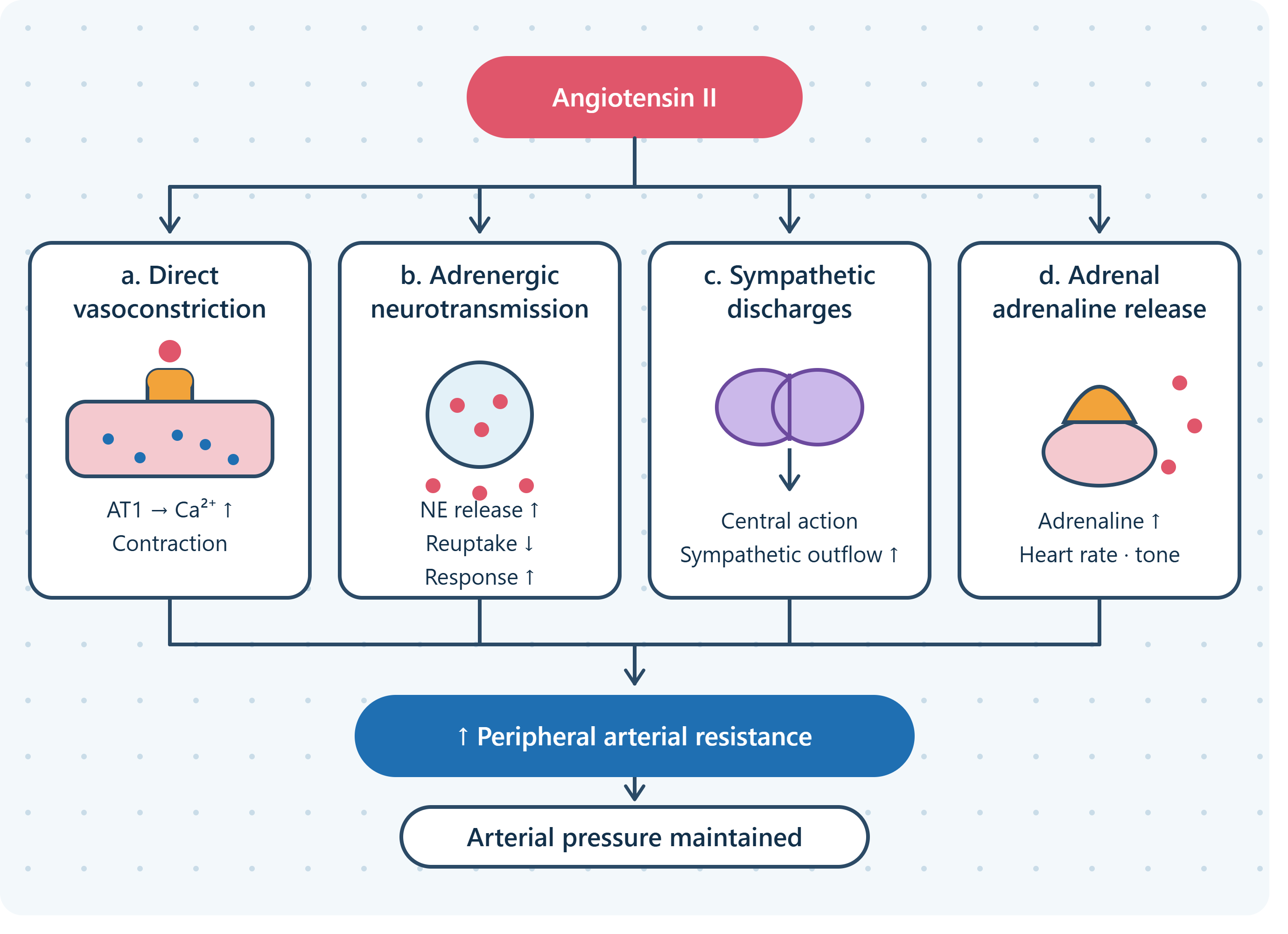
4-1. Mechanisms Responsible for the Increase in Peripheral Arterial Resistance Induced by Angiotensin II
The increase in peripheral arterial resistance induced by angiotensin II results from several complementary mechanisms involving direct vascular contraction, sympathetic nervous system stimulation, and adrenal catecholamine secretion.
a. Direct vasoconstriction
Angiotensin II produces a generalized vasoconstrictor effect by stimulating AT1 receptors located on vascular smooth muscle cells. This activation increases intracellular calcium concentrations and promotes smooth muscle contraction.
Although vasoconstriction occurs throughout the systemic circulation, its effects are particularly pronounced at the renal level. Renal vasoconstriction contributes to the regulation of renal blood flow and influences glomerular hemodynamics.
The resulting increase in vascular resistance plays an important role in maintaining arterial pressure, particularly during conditions associated with reduced circulating blood volume or decreased renal perfusion.
b. Increase in adrenergic neurotransmission
Angiotensin II enhances sympathetic neurotransmission through several mechanisms that increase the availability and vascular effects of norepinephrine.
This phenomenon is linked to:
-
Increased release of norepinephrine from sympathetic nerve endings, enhancing adrenergic stimulation of vascular smooth muscle.
-
Inhibition of norepinephrine reuptake in sympathetic nerve endings, prolonging the availability and duration of its action.
-
An increase in the vascular response to norepinephrine, resulting in greater vasoconstriction for a given level of sympathetic stimulation.
Through these mechanisms, angiotensin II amplifies the effects of the sympathetic nervous system and contributes to the maintenance of elevated peripheral vascular resistance.
c. Increase in sympathetic discharges
Angiotensin II also increases sympathetic nervous system activity through a direct effect on the central nervous system.
By acting on central regulatory structures involved in cardiovascular control, angiotensin II can stimulate sympathetic discharges and influence autonomic regulation of arterial pressure.
The resulting increase in sympathetic outflow contributes to vascular constriction, cardiac stimulation, and the enhancement of other neurohormonal responses involved in blood pressure regulation.
d. Release of adrenaline from the adrenal medulla
Angiotensin II stimulates catecholamine secretion from the adrenal medulla, particularly adrenaline.
Adrenaline acts on adrenergic receptors located in different cardiovascular tissues. Depending on the receptor subtype and tissue involved, it can influence heart rate, myocardial contractility, and vascular tone.
The release of adrenaline therefore contributes to the overall cardiovascular response associated with renin-angiotensin system activation.
Together, these mechanisms explain the powerful vasopressor action of angiotensin II and its ability to regulate peripheral arterial resistance through both direct vascular and neurohumoral pathways.
4-2. Renal Effects of Angiotensin II
Angiotensin II has important renal effects, particularly concerning sodium and potassium homeostasis, renal blood flow, and glomerular filtration.
It decreases urinary sodium (Na⁺) excretion and increases urinary potassium (K⁺) excretion, mainly through its effects on renal tubular transport mechanisms and aldosterone secretion.
These actions are essential for regulating extracellular fluid volume, maintaining electrolyte balance, and adapting renal function to changes in sodium intake and circulating blood volume.
The mechanisms responsible for the modification of renal function by angiotensin II include the following:
a. Direct increase in renal sodium reabsorption
Angiotensin II directly stimulates sodium reabsorption along different segments of the nephron.
At the proximal tubule, it increases sodium reabsorption through stimulation of sodium transport mechanisms, particularly the sodium/hydrogen exchanger (NHE3). This process facilitates the movement of sodium from tubular fluid into renal tubular cells and subsequently into the circulation.
Angiotensin II also influences sodium chloride transport in the ascending limb of the loop of Henle, including regulation of the sodium-potassium-two chloride cotransporter (NKCC2).
These effects contribute to the reduction of urinary sodium losses and help maintain extracellular fluid volume.
In healthy subjects, this regulatory mechanism helps preserve relatively stable arterial blood pressure despite substantial variations in dietary sodium intake.
For example, during low sodium intake, renin secretion increases. The resulting rise in angiotensin II stimulates renal sodium reabsorption, helping the body conserve sodium and maintain circulating blood volume.
b. Release of aldosterone from the adrenal cortex
Angiotensin II stimulates aldosterone secretion from the zona glomerulosa of the adrenal cortex.
This effect can occur at angiotensin II concentrations that do not necessarily produce significant systemic vasoconstriction, demonstrating the sensitivity of the adrenal hormonal response.
Aldosterone acts primarily on the distal nephron, particularly the late distal tubule and collecting duct, where it increases sodium reabsorption and promotes potassium secretion.
At the cellular level, aldosterone regulates the expression and activity of epithelial sodium channels (ENaC) and sodium-potassium ATPase pumps. These mechanisms facilitate sodium retention and potassium elimination in the urine.
As a result, aldosterone contributes to the maintenance of extracellular fluid volume, circulating blood volume, and arterial pressure.
c. Decrease in renal blood flow
Angiotensin II decreases renal blood flow through its direct vasoconstrictor action on the renal vascular system.
This effect is mediated mainly by AT1 receptors located on renal vascular smooth muscle cells. The resulting vasoconstriction increases renal vascular resistance and modifies the distribution of blood flow within the kidney.
In addition to its direct renal effects, angiotensin II indirectly reduces renal perfusion through increased systemic sympathetic activity and stimulation of intrarenal sympathetic mechanisms.
The magnitude of the reduction in renal blood flow depends on angiotensin II concentration, the physiological condition of the individual, and the degree of activation of other renal regulatory mechanisms.
d. Variable effect on the glomerular filtration rate
The effect of angiotensin II on the glomerular filtration rate (GFR) is complex because its vasoconstrictor action affects both the afferent and efferent arterioles of the glomerulus.
The afferent arteriole carries blood into the glomerular capillaries, whereas the efferent arteriole carries blood away from the glomerulus.
Contraction of the afferent arteriole decreases blood flow entering the glomerulus and tends to reduce glomerular capillary pressure. In contrast, contraction of the efferent arteriole increases resistance to blood leaving the glomerulus and can help maintain glomerular capillary hydrostatic pressure.
These opposing effects explain why the overall influence of angiotensin II on GFR is variable and depends on the relative degree of afferent and efferent arteriolar constriction.
In healthy subjects, the resulting effect is relatively neutral on the glomerular filtration rate under many physiological conditions. The preferential effect on the efferent arteriole helps preserve filtration pressure when renal perfusion is reduced.
In renovascular hypertension, the effect on the efferent arteriole may predominate, allowing angiotensin II to increase or maintain glomerular filtration despite reduced renal perfusion.
This mechanism also explains why sudden inhibition of the renin-angiotensin system can lead to acute renal failure in patients with bilateral renal artery stenosis.
In this pathological condition, renal perfusion is already compromised because of narrowing of both renal arteries. Glomerular filtration becomes particularly dependent on angiotensin II-mediated efferent arteriolar constriction.
Consequently, abrupt inhibition of angiotensin II production or action can cause efferent arteriolar dilation, a reduction in glomerular capillary pressure, and a significant decrease in GFR.
More generally, when renal perfusion pressure is low, angiotensin II helps preserve an adequate glomerular filtration rate by contracting the efferent arteriole of the glomerulus. This compensatory mechanism is essential for maintaining renal filtration during conditions of reduced circulating volume or impaired renal blood flow.
The renal actions of angiotensin II therefore illustrate the central role of the renin-angiotensin system in maintaining sodium balance, regulating extracellular fluid volume, and preserving glomerular filtration under changing physiological conditions.

Figure 1. The renin-angiotensin system and its main physiological components.