Feiyuebio
Rabbit TNF-α(Tumor Necrosis Factor Alpha) ELISA Kit
- SKU:
- FY-ERB4366
- Weight:
- 0 KGS
- Shipping:
- Calculated at Checkout
Description
TNF-α(Tumor Necrosis Factor Alpha) Introduction
Tumor necrosis factor (TNF, cachexin, or cachectin; often called tumor necrosis factor alpha or TNF-α) is an adipokine and a cytokine. TNF is a member of the TNF superfamily, which consists of various transmembrane proteins with a homologous TNF domain.TNF was thought to be produced primarily by macrophages, but it is produced also by a broad variety of cell types including lymphoid cells, mast cells, endothelial cells, cardiac myocytes, adipose tissue, fibroblasts, and neurons.[unreliable medical source? Large amounts of TNF are released in response to lipopolysaccharide, other bacterial products, and interleukin-1 (IL-1). In the skin, mast cells appear to be the predominant source of pre-formed TNF, which can be released upon inflammatory stimulus. In the brain TNF can protect against excitotoxicity. TNF strengthens synapses. TNF in neurons promotes their survival, whereas TNF in macrophages and microglia results in neurotoxins that induce apoptosis.TNF-α and IL-6 concentrations are elevated in obesity. Monoclonal antibody against TNF-α is associated with increases rather than decreases in obesity, indicating that inflammation is the result, rather than the cause, of obesity. TNF and IL-6 are the most prominent cytokines predicting COVID-19 severity and death.
Rabbit TNF-α(Tumor Necrosis Factor Alpha) ELISA Kit Test method
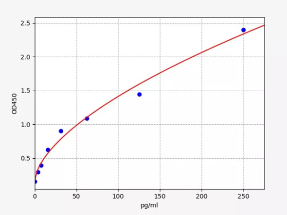
This kit uses sandwich ELISA to detect the concentration of TNF-α(Tumor Necrosis Factor Alpha) The microtiter plate provided in this kit has been pre-coated with an antibody specific to TNF-α(Tumor Necrosis Factor Alpha). Standards or samples are then added to the appropriate microtiter plate wells with a biotin-conjugated antibody specific to TNF-α(Tumor Necrosis Factor Alpha). Next, Avidin conjugated to Horseradish Peroxidase (HRP) is added to each microplate well and incubated. After TMB substrate solution is added, only those wells that contain of TNF-α, biotin-conjugated antibody and enzyme-conjugated Avidin will exhibit a change in color. The enzyme-substrate reaction is terminated by the addition of sulphuric acid solution and the color change is measured spectrophotometrically at a wavelength of 450nm ± 10nm. The concentration of TNF-α(Tumor Necrosis Factor Alpha) in the samples is then determined by comparing the OD of the samples to the standard curve.
Reference:
1, Old LJ (1985). "Tumor necrosis factor (TNF)". Science. 230 (4726): 630–2. Bibcode:1985Sci...230..630O. doi:10.1126/science.2413547. PMID 2413547.
2, Nedwin GE, Naylor SL, Sakaguchi AY, Smith D, Jarrett-Nedwin J, Pennica D, Goeddel DV, Gray PW (1985). "Human lymphotoxin and tumor necrosis factor genes: structure, homology and chromosomal localization". Nucleic Acids Res. 13 (17): 6361–73. doi:10.1093/nar/13.17.6361. PMC 321958. PMID 2995927.
3, Kriegler M, Perez C, DeFay K, Albert I, Lu SD (1988). "A novel form of TNF/cachectin is a cell surface cytotoxic transmembrane protein: ramifications for the complex physiology of TNF". Cell. 53 (1): 45–53. doi:10.1016/0092-8674(88)90486-2. PMID 3349526. S2CID 31789769.
Additional Information
Size: |
96T |
Alias: |
TNF-α ELISA Kit |
Detection Method: |
Sandwich ELISA, Double Antibody |
Application: |
TNF-α ELISA Kit allows for the in vitro quantitative determination of TNF-α concentrations in serum, plasma, tissue homogenates and other biological fluids. |
Storage: |
2-8 ℃ for 6 months |
Sensitivity: |
<9.375 pg/ml |
Species: |
Rabbit |
Uniprot: |
P04924 |
CV %(): |
Intra-Assay: CV<8% Inter-Assay: CV<10% |


