Feiyuebio
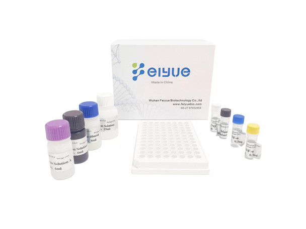
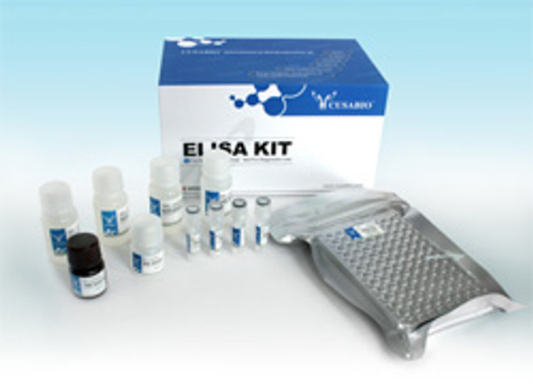
Rat TNF-α(Tumor Necrosis Factor Alpha) ELISA Kit
- SKU:
- FY-ER4365
- Weight:
- 0 KGS
- Shipping:
- Calculated at Checkout
Description
Rat TNF-α(Tumor Necrosis Factor Alpha) ELISA Kit Basic Information
|
Product Name |
Rat TNF-α(Tumor Necrosis Factor Alpha) ELISA Kit |
|
Catalog NO. |
FY-ER4365 |
|
Alias |
TNF-α ELISA Kit, Serum Amyloid A ELISA Kit |
|
Application |
TNF-α ELISA Kit allows for the in vitro quantitative determination of TNF-α concentrations in serum, plasma, tissue homogenates and other biological fluids. |
|
Size |
48T, 96T |
|
Detection Method |
Sandwich ELISA, Double Antibody |
|
Storage |
2-8 ℃ for 6 months |
|
Sensitivity |
< 2.344pg/ml |
|
Species |
Rat |
|
UniProt ID |
P16599 |
|
CV %() |
Intra-Assay: CV<8% |
|
Note |
For Research Use Only |
Feiyue's Rat TNF-α is an ELISA reagent for detection of TNF-α in Rat serum, plasma or cell with sensitivity, specificity and consistency.
TNF-α(Tumor necrosis factor alpha) Introduciton
Tumor necrosis factor (TNF, cachexin, or cachectin; often called tumor necrosis factor alpha or TNF-α) is an adipokine and a cytokine. TNF is a member of the TNF superfamily, which consists of various transmembrane proteins with a homologous TNF domain. TNF was thought to be produced primarily by macrophages, but it is produced also by a broad variety of cell types including lymphoid cells, mast cells, endothelial cells, cardiac myocytes, adipose tissue, fibroblasts, and neurons.[unreliable medical source?] Large amounts of TNF are released in response to lipopolysaccharide, other bacterial products, and interleukin-1 (IL-1). In the skin, mast cells appear to be the predominant source of pre-formed TNF, which can be released upon inflammatory stimulus (e.g., LPS).TNF has been shown to interact with TNFRSF1A.
Rat TNF-α(Tumor Necrosis Factor Alpha) ELISA Kit Test method
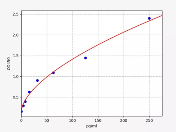
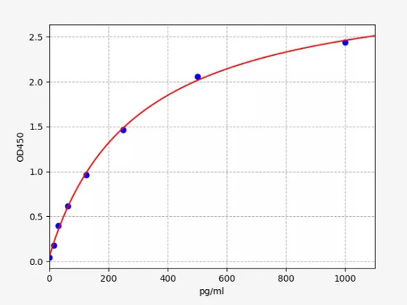
This kit uses sandwich ELISA to detect the concentration of TNF-α(Tumor necrosis factor alpha)) The microtiter plate provided in this kit has been pre-coated with an antibody specific to TNF-α(Tumor necrosis factor alpha)). Standards or samples are then added to the appropriate microtiter plate wells with a biotin-conjugated antibody specific to TNF-α(Tumor necrosis factor alpha)). Next, Avidin conjugated to Horseradish Peroxidase (HRP) is added to each microplate well and incubated. After TMB substrate solution is added, only those wells that contain of TNF-α, biotin-conjugated antibody and enzyme-conjugated Avidin will exhibit a change in color. The enzyme-substrate reaction is terminated by the addition of sulphuric acid solution and the color change is measured spectrophotometrically at a wavelength of 450nm ± 10nm. The concentration of TNF-α(Tumor necrosis factor alpha)) in the samples is then determined by comparing the OD of the samples to the standard curve.
Reference:
1, Heir R, Stellwagen D (2020). "TNF-Mediated Homeostatic Synaptic Plasticity: From in vitro to in vivo Models". Frontiers in Cellular Neuroscience. 14: 565841. doi:10.3389/fncel.2020.565841. PMC 7556297. PMID 33192311.
2, Gough P, Myles IA (2020). "Tumor Necrosis Factor Receptors: Pleiotropic Signaling Complexes and Their Differential Effects". Frontiers in Immunology. 11: 585880. doi:10.3389/fimmu.2020.585880. PMC 7723893. PMID 33324405.
Additional Information
Size: |
96T |
Reactivity: |
Rat |
Assay range: |
3.906-250pg/ml |
sensitivity: |
2.344pg/ml |

