
Siemens
Siemens Clinitest 20 swabs

- SKU:
- 1574745
- Availability:
- Immediate shippement after your order. Less than 24h delivery
- Weight:
- 1 KGS
- Shipping:
- €35.00 (Fixed Shipping Cost)
Bulk discount rates
Below are the available bulk discount rates for each individual item when you purchase a certain amount
| Buy 10 - 47 | and get 5% off |
| Buy 48 or above | and get 10% off |

Description
Siemen shallow nasal swab, 20 tests
INTENDED USE
The CLINITEST® Rapid Antigen Test is an IVD immunochromatographic assay for the qualitative detection of
nucleocapsid protein antigen in direct nasopharyngeal (NP) swab or nasal swab specimens directly from individuals who are suspected.
It is intended to aid in the rapid testing. Negative results from patients with symptom onset beyond ten days should be
treated as presumptive and confirmation with a molecular assay, if necessary, for patient management, may be performed.
The CLINITEST Rapid Antigen Test does not differentiate between Omicron and Delta but detects very well Omicron.
SUMMARY AND EXPLANATION
The Delta incubation period is 1 to 14 days, mostly 3 to 7 days. Omicron 2 to 6 days only. The main manifestations include fever, fatigue, and dry cough. Nasal congestion, runny nose, sore throat, myalgia, and diarrhea are found in a few cases.
This test is for detection of nucleocapsid protein antigen. Antigen is generally detectable in upper respiratory specimens during the acute phase of infection. Rapid detection will help you to control control the disease more efficiently and effectively.
PRINCIPLE OF THE TEST
The CLINITEST Self Antigen Test is an immunochromatographic membrane assay that uses highly sensitive monoclonal antibodies to detect nucleocapsid protein in direct nasopharyngeal (NP) swab or nasal swab.
The test strip is composed of the following parts: namely sample pad, reagent pad, reaction membrane, and absorbing pad. The reagent pad contains the colloidal-gold conjugated with the monoclonal antibodies against the nucleocapsid protein.
The reaction membrane contains the secondary antibodies for nucleocapsid protein. The whole strip is fixed inside a plastic device. When the sample is added into the sample well, conjugates dried in the reagent pad are dissolved and migrate along with the sample. If the omicron nucleocapsid antigen is present in the sample, a complex forms between the anti-SARS-2 conjugate and the virus will be captured by the specific anti-S monoclonal antibodies coated on the test line region (T). Absence of the test line (T) suggests a negative result. To serve as a procedural control, a red line will always appear in the control line region (C) indicating that proper volume of sample has been added and membrane wicking has occurred.
MATERIALS PROVIDED
- 20 Test Cassettes
- 2 Extraction Buffer Vials
- 20 Sterile Swabs
- 20 Extraction Tubes and Tips
- 1 Workstation
- 1 Package Insert
MATERIALS REQUIRED BUT NOT PROVIDED
1. Clock, timer, or stopwatch
WARNINGS AND PRECAUTIONS
1. For in vitro diagnostic use only.
2. The test device should remain in the sealed pouch until use.
3. Do not use kit past its expiration date.
4. Swabs, tubes, and test devices are for single use only.
5. Solutions that contain sodium azide may react explosively with lead or copper plumbing. Use large quantities of water to flush
discarded solutions down a sink.
6. Do not interchange or mix components from different kit lots.
7. Testing should only be performed using the swabs provided within the kit.
8. To obtain accurate results, do not use visually bloody or overly viscous samples.
9. Wear appropriate personal protection equipment and gloves when running each test and handling patient specimens. Change
gloves between handling of specimens suspected of COVID-19.
10. Specimens must be processed as indicated in the SPECIMEN COLLECTION and SAMPLE PREPARATION PROCEDURE
sections of this Product Insert. Failure to follow the instructions for use can result in inaccurate results.
11. Proper laboratory safety techniques should be followed at all times when working with SARS-CoV-2 patient samples, swabs used Test Strips and used extraction buffer vials may be potentially infectious. Proper handling and disposal methods
should be established by the laboratory in accordance with local regulatory requirements.
12. Inadequate or inappropriate specimen collection and storage can adversely affect results.
13. Humidity and temperature can adversely affect results.
14. Dispose of test device and materials as biohazardous waste in accordance with federal, state, and local requirements.
STORAGE AND STABILITY
1. The kit can be stored at room temperature or refrigerated (2-30°C).
2. Do not freeze any of the test kit components.
3. Do not use test device and reagents after expiration date.
4. Test devices that have been outside of the sealed pouch for more than 1 hour should be discarded.
5. Close the kit box and secure its contents when not in use.
SPECIMEN COLLECTION
1. Nasopharyngeal Swab
1) Using the sterile swab provided in the kit, carefully insert the swab in the patient’s nostril.
2) Swab over the surface of the posterior nasopharynx and rotate the swab several times.
3) Withdraw the swab from the nasal cavity. The specimen is now ready for preparation using the extraction buffer provided in
the test kit.
2. Nasal Swab
1) Using the sterile swab provided in the kit, carefully insert the swab into one nostril of the patient. The swab tip should be
inserted up to 2-4 cm until resistance is met.
2) Roll the swab 5 times along the mucosa inside the nostril to ensure that both mucus and cells are collected.
3) Using the same swab, repeat this process for the other nostril to ensure that an adequate sample is collected from both nasal cavities.
4) Withdraw the swab from the nasal cavity. The specimen is now ready for preparation using the extraction buffer provided in
the test kit.
SAMPLE PREPARATION PROCEDURE
1. Insert the test extraction tube into the workstation provided in the kit. Make sure that the tube is standing upright and reaches
the bottom of the workstation.
2. Add 0.3 mL (approximately 10 drops) of the sample extraction buffer into the extraction tube.
CLINITEST®Rapid Antigen Test
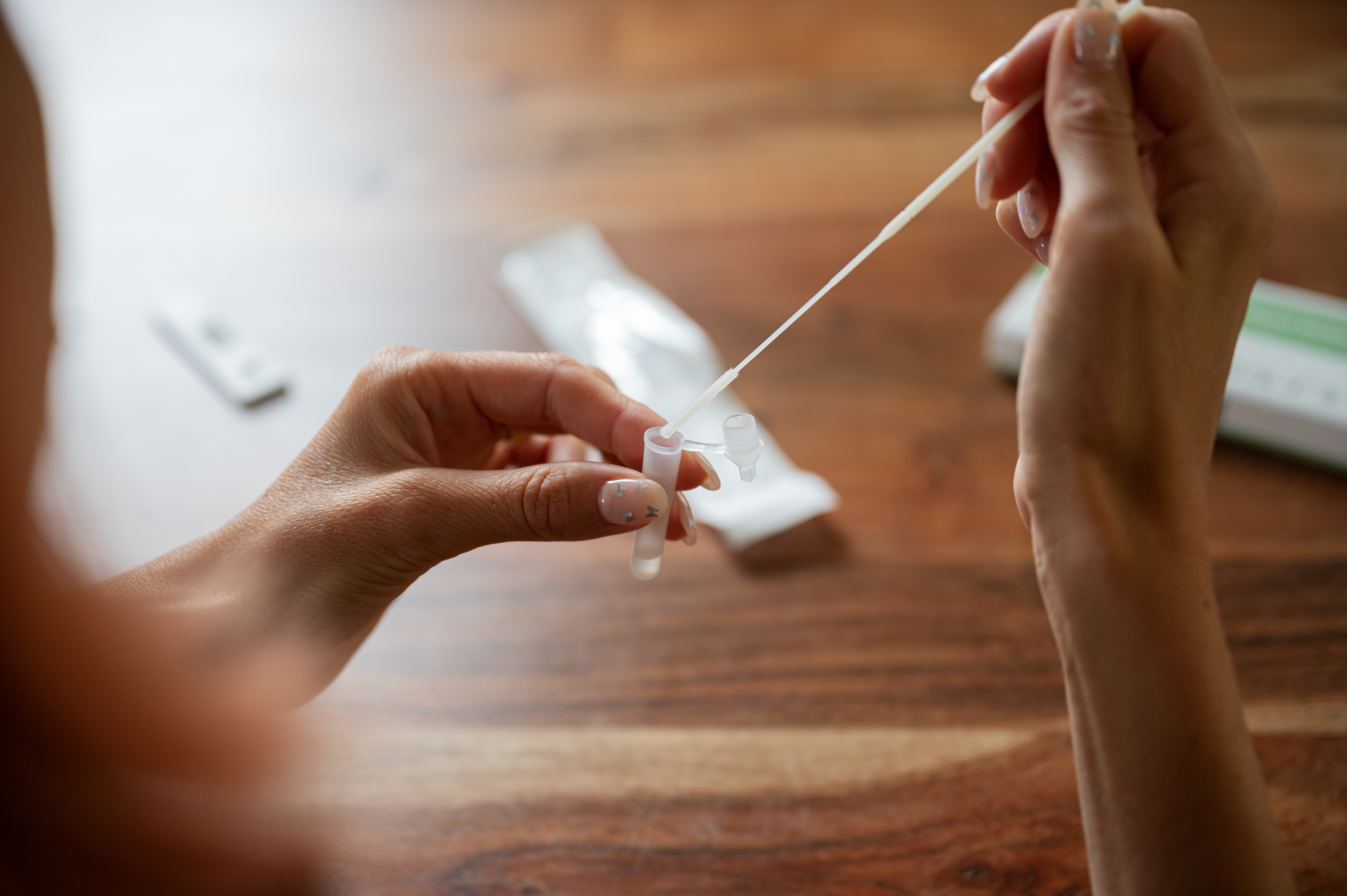
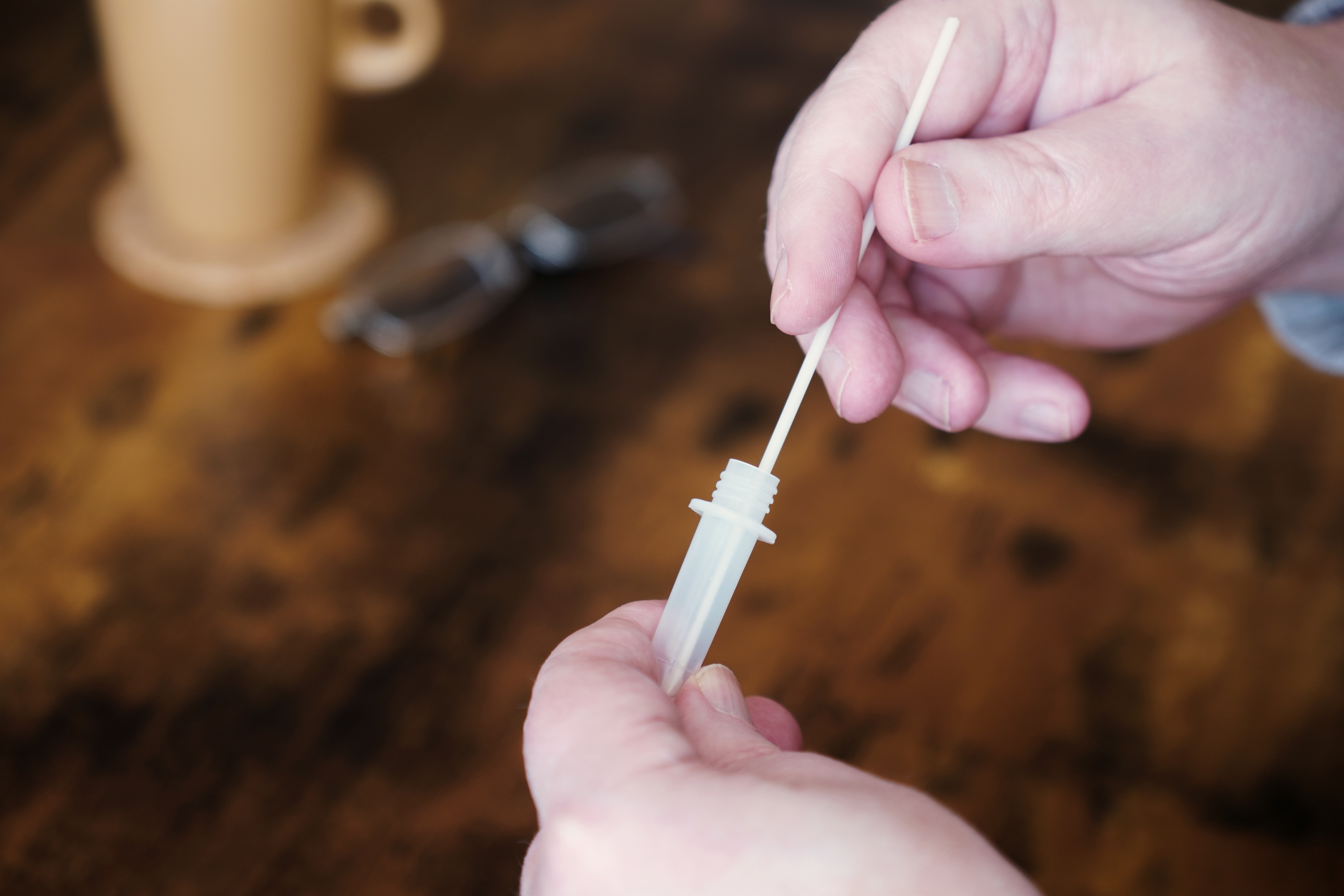
3. Insert the swab into the extraction tube which contains 0.3 mL of the extraction buffer.
4. Roll the swab at least 6 times while pressing the head against the bottom and side of the extraction tube.
5. Leave the swab in the extraction tube for 1 minute.
6. Squeeze the tube several times from the outside to immerse the swab. Remove the swab.
SPECIMEN TRANSPORT AND STORAGE
Do not return the sterile swab to the original paper packaging.
Specimen should be tested immediately after collection. If immediate testing of specimen is not possible, insert the swab
into an unused general-purpose plastic tube. Ensure the breakpoint swab is level with the tube opening. Bend the swab
shaft at a 180 degrees angle to break it off at the breaking point. You may need to gently rotate the swab shaft to complete
the breakage. Ensure the swab fits within the plastic tube and secure a tight seal. The specimen should be disposed and
recollected for retesting if untested for longer than 1 hour.
TEST PROCEDURE
Allow the test device, test sample and buffer to equilibrate to room temperature (15-30°C) prior to testing.
1. Just prior to testing remove the test device from the sealed pouch and place it on a flat surface.
2. Push the nozzle which contains the filter onto the extraction tube. Ensure the nozzle has a tight fit.
3. Hold the extraction tube vertically and add 4 drops (approximately 100 μL) of test sample solution tube into the sample well.
4. Start the timer.
5. Read the results at 15 minutes. Do not interpret the result after 20 minutes.
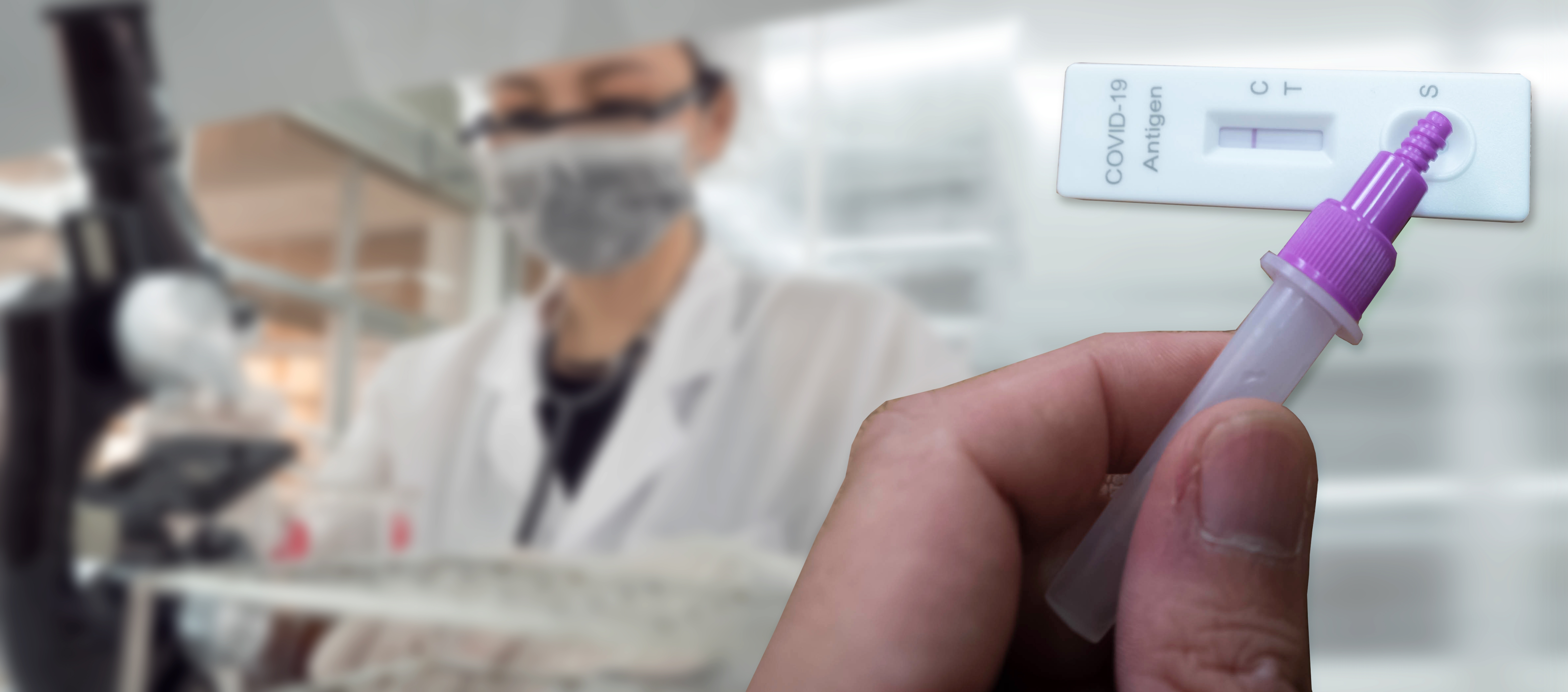
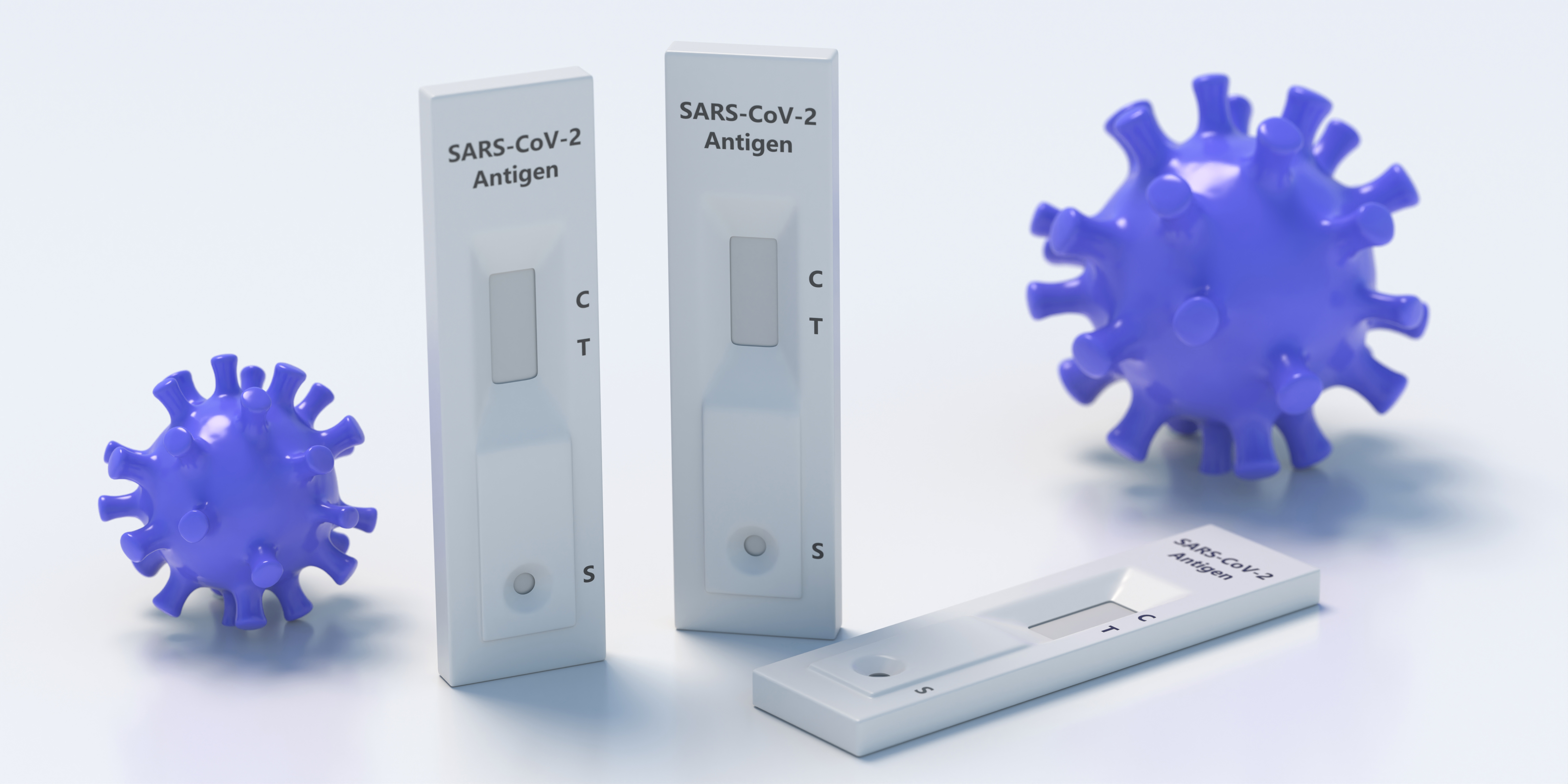
INTERPRETATION OF RESULTS
1. POSITIVE:
The presence of two lines as control line (C) and test line (T) within the result window indicates a positive result.
2. NEGATIVE:
The presence of only control line (C) within the result window indicates a negative result.
3. INVALID:
If the control line (C) is not visible within the result window after performing the test, the result is considered invalid. Some causes
of invalid results are because of not following the directions correctly or the test may have deteriorated beyond the expiration date.
It is recommended that the specimen be re-tested using a new test.
NOTE:
1.The intensity of color in the test line region (T) may vary depending on the concentration of analyses present in the specimen.
Therefore, any shade of color in the test line region (T) should be considered positive. This is a qualitative test only and cannot
determine the concentration of analytes in the specimen.
2.Insufficient specimen volume, incorrect operating procedure or expired tests are the most likely reasons for control band failure.
QUALITY CONTROL
A procedural control is included in the test. A red line appearing in the control line region (C) is the internal procedural control. It
confirms sufficient specimen volume and correct procedural technique. Control standards are not supplied with this test. However,
it is recommended that positive and negative controls are sourced from a local competent authority and tested as a good laboratory
practice, to confirm the test procedure and verify the test performance.
LIMITATIONS
1. The etiology of respiratory infection caused by microorganisms other than SARS-CoV-2 will not be established with this test.
The CLINITEST Rapid COVID-19 Antigen Test can detect both viable and non-viable SARS-CoV-2. The performance of the
CLINITEST Rapid COVID-19 Antigen Test depends on antigen load and may not correlate with viral culture results performed
on the same specimen.
2. Failure to follow the Test Procedure may adversely affect test performance and/or invalidate the test result.
3. If the test result is negative and clinical symptoms persist, additional testing using other clinical methods is recommended. A
negative result does not at any time rule out the presence of SARS-CoV-2 antigens in specimen, as they may be present below
the minimum detection level of the test or if the sample was collected or transported improperly.
4. As with all diagnostic tests, a confirmed diagnosis should only be made by a physician after all clinical and laboratory findings
have been evaluated.
5. Positive test results do not rule out co-infections with other pathogens.
6. Positive test results do not differentiate between SARS-CoV and SARS-CoV-2.
7. The amount of antigen in a sample may decrease as the duration of illness increases. Specimens collected after day 10 of
illness are more likely to be negative compared to a RT-PCR assay.
8. Negative results from patients with symptom onset beyond ten days, should be treated as presumptive and confirmation with a
molecular assay, if necessary, for patient management, may be performed.
9. Negative results do not rule out SARS-CoV-2 infection and should not be used as the sole basis for treatment or patient
management decisions, including infection control decisions.

PERFORMANCE CHARACTERISTICS
1. Clinical Sensitivity, Specificity and Accuracy
Nasopharyngeal Swab
Clinical Performance of the CLINITEST Rapid COVID-19 Antigen Test was evaluated by being involved in 7 sites within the US
where patients were enrolled and tested. Testing was performed by 24 Healthcare Workers that were not familiar with the testing
procedure. A total of 865 fresh nasopharyngeal swab samples was collected and tested, which includes 119 positive samples and
746 negative samples.
The CLINITEST Rapid COVID-19 Antigen Test results were compared to USFDA Emergency Use Authorized
RT-PCR assays for SARS-CoV-2 in nasopharyngeal swab specimens.
Overall study results are shown in Table 1.
Table 1: CLINITEST Rapid COVID-19 Antigen Test (Nasopharyngeal Swab) vs PCR
Method PCR
Total Results
CLINITEST Rapid
C19 Antigen Test
(Nasopharyngeal Swab)
Results Positive Negative
Positive 117 3 120
Negative 2 743 745
Total 119 746 865
Relative Sensitivity: 98.32% (95% Cl* 94.06% to 99.80%) *Confidence Intervals
Relative Specificity: 99.60% (95% CI* 98.83.03% to 99.92%)
Accuracy: 99.42% (95%CI* 98.66% to 99.81%)
Nasal Swab
A total of 237 fresh nasal swab samples was collected and tested, which includes 109 positive samples and 128 negative samples.
The CLINITEST Rapid Antigen Test results were compared to results of USFDA Emergency Use Authorized RT-PCR
assays for SARS-CoV-2 in nasopharyngeal swab specimens. Overall study results are shown in Table 2.
Table 2: CLINITEST Rapid COVID-19 Antigen Test (Nasal Swab) vs PCR
Method PCR
Total Results
CLINITEST Rapid C#19 Antigen Test
(Nasal Swab)
Results Positive Negative
Positive 106 0 106
Negative 3 128 131
Total 109 128 237
Relative Sensitivity: 97.25% (95% Cl*: 92.17% to 99.43%) *Confidence Intervals
Relative Specificity: 100% (95% CI*: 97.16% to 100%)
Accuracy: 98.73% (95%CI*: 96.35% to 99.74%)
2. Limit of Detection (LOD)
LOD studies determine the lowest detectable concentration of SARS-CoV-2 at which approximately 95% of all (true positive)
replicates test positive. Heat inactivated SARS-CoV-2 virus, with a stock concentration of 4.6 x 10⁵ TCID50 / mL, was spiked into
negative specimen and serially diluted. Each dilution was ran in triplicate on the CLINITEST Rapid COVID-19 Antigen Test. The
Limit of Detection of the CLINITEST Rapid COVID-19 Antigen Test is 1.15 x 10² TCID50 / mL (Table 3).
Table 3: Limit of Detection (LOD) Study Results
Concentration No. Positive/Total Positive Agreement
1.15 x 10² TCID50 / mL 180/180 100%
3. High Dose Hook Effect
No high dose hook effect was observed when testing up to a concentration of 4.6 x 10⁵ TCID50 / mL of heat inactivated SARS-CoV2 virus.
4. Cross Reactivity
Cross reactivity with the following organisms has been studied. Samples positive for the following organisms were found negative when tested with the CLINITEST Rapid COVID-19 Antigen Test.
Pathogens Concentration
- Respiratory syncytial virus Type A 5.5×10⁷ PFU/mLRespiratory syncytial virus Type B 2.8×10⁵ TCID50/mL
Novel influenza A H1N1 virus (2009) 1×10⁶ PFU/mL
Seasonal influenza A H1N1 virus 1×10⁵ PFU/mL
Influenza A H3N2 virus 1×10⁶ PFU/mL
Influenza A H5N1 virus 1×10⁶ PFU/mL
Influenza B Yamagata 1×10⁵ PFU/mL
Influenza B Victoria 1×10⁶ PFU/mL
Rhinovirus 1×10⁶ PFU/mL
Adenovirus 3 5×107.5 TCID50/mL
Adenovirus 7 2.8×10⁶ TCID50/mL
EV-A71 1×10⁵ PFU/mL
Mycobacterium tuberculosis 1×10³ bacteria/mL
Mumps virus 1×10⁵ PFU/mL
Human coronavirus 229E 1×10⁵ PFU/mL
Human coronavirus OC43 1×10⁵ PFU/mL
Human coronavirus NL63 1×10⁶ PFU/mL
Human coronavirus HKU1 1×10⁶ PFU/mL
Parainfluenza virus 1 7.3×10⁶ PFU/mL
Parainfluenza virus 2 1×10⁶ PFU/mL
Parainfluenza virus 3 5.8×10⁶ PFU/mL
Parainfluenza virus 4 2.6×10⁶ PFU/mL
Haemophilus influenzae 5.2×10⁶ CFU/mL
Streptococcus pyogenes 3.6×10⁶ CFU/mL
Streptococcus pneumoniae 4.2×10⁶ CFU/mL
Candida albicans 1×10⁷ CFU/mL
Bordetella pertussis 1×10⁴ bacteria/mL
Mycoplasma pneumoniae 1.2×10⁶ CFU/mL
Chlamydia pneumoniae 2.3×10⁶ IFU/mL
Legionella pneumophila 1×10⁴ bacteria/mL
Staphylococcus aureus 3.2×10⁸ CFU/mL
Staphylococcus epidermidis 2.1×10⁸ CFU/mL
5. Interfering Substance
The following substances, naturally present in respiratory specimens or that may be artificially introduced into the nasal cavity or
nasopharynx, were evaluated with the CLINITEST Rapid COVID-19 Antigen Test at the concentrations listed below and were found
not to affect test performance.
Substance Concentration
Human blood (EDTA anticoagulated) 20% (v/v)
6. Microbial Interference
To evaluate whether potential microorganisms in clinical samples interfere with the detection of CLINITEST Rapid COVID-19
Antigen Test so as to produce false negative results. Each pathogenic microorganism was tested in triplicate in the presence of
heat inactivated SARS-Cov-2 virus (2.3x10² TCID50/swab). No cross reactivity or interference was seen with the microorganisms
listed in the table below.
Microorganism Concentration
- Respiratory syncytial virus Type A 5.5×10⁷PFU/mlRespiratory syncytial virus Type B 2.8×10⁵ TCID50/mlNovel influenza A H1N1 virus (2009) 1×10⁶PFU/ml
Seasonal influenza A H1N1 virus 1×10⁵PFU/ml
Influenza A H3N2 virus 1×10⁶PFU/ml
Influenza A H5N1 virus 1×10⁶PFU/ml
Influenza B Yamagata 1×10⁵PFU/ml
Influenza B Victoria 1×10⁶PFU/ml
Rhinovirus 1×10⁶PFU/ml
Adenovirus 1 1×10⁶PFU/ml
Adenovirus 2 1×10⁵PFU/ml
Adenovirus 3 5×107.5 TCID50/ml
Adenovirus 4 1×10⁶PFU/ml
Adenovirus 5 1×10⁵PFU/ml
Adenovirus 7 2.8×10⁶ TCID50/ml
Adenovirus 55 1×10⁵PFU/ml
EV-A71 1×10⁵PFU/ml
EV-B69 1×10⁵PFU/ml
EV-C95 1×10⁵PFU/ml
EV-D70 1×10⁵PFU/ml
Mycobacterium tuberculosis 1×10³ bacterium/ml
Mumps virus 1×10⁵PFU/ml
Varicella zoster virus 1×10⁶PFU/ml
Human coronavirus 229E 1×10⁵PFU/ml
Human coronavirus OC43 1×10⁵PFU/ml
Human coronavirus NL63 1×10⁶PFU/ml
Human coronavirus HKU1 1×10⁶PFU/ml
Human Metapneumovirus (hMPV) 1×10⁶PFU/ml
Parainfluenza virus 1 7.3×10⁶PFU/ml
Parainfluenza virus 2 1×10⁶PFU/ml
Parainfluenza virus 3 5.8×10⁶PFU/ml
Parainfluenza virus 4 2.6×10⁶PFU/ml
Haemophilus influenzae 5.2×10⁶ CFU/ml
Streptococcus pyogenes 3.6×10⁶ CFU/ml
Streptococcus agalactiae 7.9×10⁷ CFU/ml
Streptococcus pneumoniae 4.2×10⁶ CFU/ml
Candida albicans 1×10⁷ CFU/ml
Bordetella pertussis 1×10⁴ bacterium/ml
Mycoplasma pneumoniae 1.2×10⁶ CFU/ml
Chlamydia pneumoniae 2.3×10⁶ IFU/ml
Legionella pneumophila 1×10⁴ bacterium/ml
Pooled human nasal wash N/A
Warranty Information
Related Products

Swabs kit -box of 75 swabs mixed
National Diagnostics

Swab Woodstick Plain in Tube
National Diagnostics

Surface ATP Test Swab
National Diagnostics

Surface Protein Test Swab
National Diagnostics

Swab 10ml Buffer+ViscoseTip
National Diagnostics