
Cell-Free DNA (cfDNA): Biology, Analysis, Isolation and Quality Control
Cell-Free DNA (cfDNA): Biology, Analysis, Isolation and Quality Control
A scientific overview of cell-free DNA, its biological origins, analytical challenges, sample handling, isolation methods, and quality control in molecular biology and clinical research.
1. What Is Cell-Free DNA?
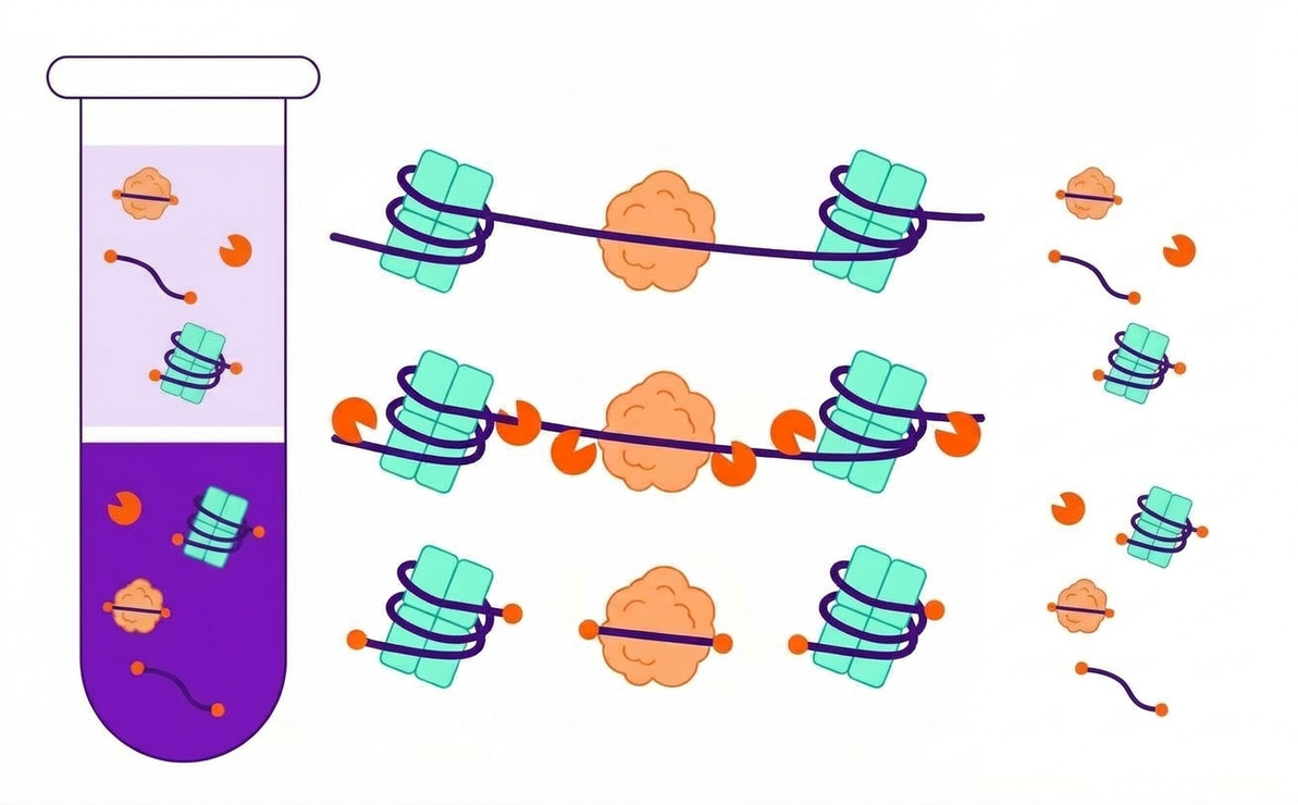
Cell-free DNA (cfDNA) consists of extracellular DNA fragments present in biological fluids such as blood plasma, serum, and urine. These fragments originate from different cell populations and are released through processes including apoptosis, necrosis, and other cellular mechanisms.
cfDNA analysis is used in oncology research, prenatal screening, transplantation research, infectious disease studies, and biomarker discovery. Its concentration and molecular characteristics can vary with physiological conditions, disease processes, and sample handling.
2. cfDNA Biology
Sources and Release Mechanisms
Apoptosis: Programmed cell death can generate DNA fragments associated with nucleosome organization.
Necrosis: Cell injury and uncontrolled cell death can release DNA fragments with heterogeneous sizes.
Active release: Certain cells may release DNA-containing material through extracellular vesicles or other biological processes.
Fragment Size and Stability
Many plasma cfDNA fragments associated with nucleosomes are approximately 160–180 base pairs long, although shorter and longer fragments can also occur. Fragment profiles depend on biological origin, degradation, and sample processing.
3. Challenges in cfDNA Analysis
| Challenge | Impact | Quality consideration |
|---|---|---|
| Low DNA concentration | May reduce detection sensitivity. | Use suitable sensitive analytical methods. |
| Genomic DNA contamination | Can alter DNA concentration and fragment profiles. | Minimize cellular disruption during processing. |
| DNA degradation | Can affect target recovery. | Control storage and processing conditions. |
| Analytical errors | Can cause false-positive or false-negative findings. | Include validated controls. |
4. Preanalytical Handling and Stability
Blood, Serum, and Plasma
Plasma is commonly used for cfDNA analysis because blood coagulation is avoided. Serum preparation may release additional genomic DNA from blood cells, potentially increasing background DNA levels.
- Use an appropriate blood collection tube.
- Process samples according to a validated protocol.
- Separate plasma carefully from blood cells.
- Minimize unnecessary sample transfers and freeze-thaw cycles.
- Follow validated storage conditions.
Urine
Urinary cfDNA is investigated in non-invasive molecular research. Its concentration and integrity may be affected by pH, nuclease activity, microbial growth, and processing delays. Collection and stabilization procedures should be appropriate for the intended analytical application.
5. cfDNA Isolation Methods
Silica-Based Extraction
DNA binds to silica membranes or particles under appropriate chemical conditions. Contaminants are removed during washing, and DNA is recovered in a suitable elution buffer.
Magnetic Bead-Based Extraction
DNA binds to functionalized magnetic particles, allowing separation from contaminants using a magnetic field. These methods can support manual or automated workflows.
General Isolation Workflow
1. Sample collection: Obtain the appropriate biological specimen.
2. Sample processing: Separate cellular components where required.
3. DNA extraction: Bind, wash, and recover cfDNA using a validated method.
4. Quality control: Assess concentration and fragment distribution when appropriate.
5. Downstream analysis: Perform qPCR, digital PCR, or sequencing as required.
6. cfDNA Quality Control
DNA Quantification
Fluorometric assays can quantify low concentrations of DNA. qPCR and digital PCR can measure specific amplifiable targets. Spectrophotometric measurements should be interpreted cautiously when DNA concentrations are very low.
Fragment Size Distribution
Capillary electrophoresis can characterize DNA fragment lengths and help identify potential contamination by high-molecular-weight genomic DNA.
Purity and Functional Assessment
Residual extraction reagents, salts, and proteins may interfere with downstream enzymatic reactions. Internal controls and validated amplification assays can help assess whether the extracted DNA is suitable for the intended application.
7. Downstream Applications
Quantitative PCR: Measures selected DNA sequences and supports targeted quantification.
Digital PCR: Estimates target concentration using partitioned amplification reactions.
Next-generation sequencing: Enables broader investigation of genetic variants and other molecular characteristics.
8. Best Practices for Reliable Results
- Standardize sample collection, processing, and storage.
- Select extraction methods compatible with the biological matrix.
- Minimize contamination from cellular genomic DNA.
- Use sensitive quantification methods suitable for low-input samples.
- Include appropriate extraction and analytical controls.
- Document sample handling and quality control results.
- Interpret results according to the method's analytical limitations.
Conclusion
Cell-free DNA is an important molecular analyte for investigating biological processes through minimally invasive sampling. Reliable cfDNA analysis depends on understanding its biological origins, controlling preanalytical variables, selecting suitable isolation methods, and implementing rigorous quality control.
Recent Posts
-
DNA Analysis Using Analytical Gels
DNA Analysis Using Analytical Gels Principles, Methods, Interpretation, and Applications of Gel El …20th Oct 2026 -
Cell-Free DNA (cfDNA): Biology, Analysis, Isolation and Quality Control
Cell-Free DNA (cfDNA): Biology, Analysis, Isolation and Quality Control A scientific overview of ce …20th Oct 2026 -
Gel Electrophoresis Equipment
Gel Electrophoresis Equipment: Types, Components, Working Principles, and Applications Introduction …19th Oct 2026