
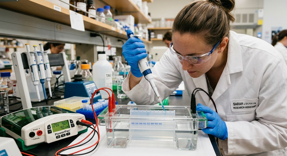
Gel Electrophoresis Equipment
Gel Electrophoresis Equipment: Types, Components, Working Principles, and Applications
Introduction
Gel electrophoresis equipment is essential in molecular biology, biotechnology, genetics, and biochemistry laboratories. This analytical technique separates DNA, RNA, or proteins according to their size, charge, and other physicochemical properties by applying an electric field across a gel matrix.
Researchers use gel electrophoresis to analyze nucleic acids, evaluate PCR products, assess DNA fragments, examine protein samples, and verify the quality of laboratory experiments. Selecting the appropriate equipment helps ensure reliable separation, reproducible results, and efficient sample analysis.
What Is Gel Electrophoresis Equipment?
Gel electrophoresis equipment refers to the laboratory instruments and accessories used to perform electrophoretic separation. A standard system typically includes an electrophoresis chamber, a gel support or casting tray, electrodes, a power supply, and a suitable buffer solution.
Depending on the application, laboratories may also require a gel documentation system, a UV or blue-light transilluminator, sample-loading accessories, and specialized staining reagents.
The equipment generates an electric field that drives charged molecules through a porous gel. Smaller molecules generally migrate more rapidly through the gel matrix than larger molecules under comparable conditions.
Main Components of Gel Electrophoresis Equipment
1. Electrophoresis Chamber
The electrophoresis chamber holds the gel and running buffer during separation. It contains electrodes that establish the electric field required to move molecules through the gel. Horizontal chambers are commonly used for DNA and RNA analysis, while vertical chambers are frequently used for protein separation.
2. Power Supply
The power supply delivers a controlled voltage or current to the electrophoresis chamber. Many laboratory power supplies allow users to adjust voltage, current, and running time according to the gel type and experimental requirements. Stable electrical conditions are important for reproducible migration and effective separation.
3. Gel Casting Tray and Comb
The casting tray provides a mold for preparing the gel. A comb is inserted into the liquid gel before it solidifies to create wells for sample loading. The number and size of the wells depend on the comb design and the volume of samples to be analyzed.
4. Electrodes and Electrical Connections
Electrodes generate the electric field inside the chamber. In conventional nucleic acid electrophoresis, negatively charged DNA migrates toward the positive electrode. Correct electrode orientation is essential because an incorrect setup can cause samples to migrate in the wrong direction.
5. Buffer Solution
The running buffer maintains a suitable ionic environment and enables electrical current to pass through the gel. Common buffers for nucleic acid electrophoresis include TAE (Tris-acetate-EDTA) and TBE (Tris-borate-EDTA). The appropriate buffer depends on the sample type, gel system, and analytical objective.
6. Gel Visualization System
After electrophoresis, separated molecules may be visualized using suitable stains and an imaging system. DNA and RNA can be detected using fluorescent stains, while proteins may be visualized using Coomassie staining or silver staining. Depending on the stain, visualization may require UV illumination, blue-light excitation, or conventional imaging.
Types of Gel Electrophoresis Equipment
Horizontal Gel Electrophoresis Systems
Horizontal electrophoresis systems are widely used for DNA and RNA analysis. They generally employ agarose gels submerged in buffer inside a horizontal chamber. Typical applications include PCR product analysis, DNA fragment separation, restriction digestion analysis, and nucleic acid quality assessment.
Vertical Gel Electrophoresis Systems
Vertical electrophoresis systems are commonly used for protein analysis. They typically use polyacrylamide gels positioned between glass plates. SDS-PAGE, or sodium dodecyl sulfate–polyacrylamide gel electrophoresis, separates denatured proteins primarily according to their molecular mass.
Agarose Gel Electrophoresis Equipment
Agarose gel systems are particularly suitable for separating DNA and RNA fragments across a range of sizes. The concentration of agarose influences the effective pore size of the gel and therefore affects separation performance. Higher agarose concentrations generally provide better resolution for smaller nucleic acid fragments, whereas lower concentrations are often used for larger fragments.
Polyacrylamide Gel Electrophoresis Equipment
Polyacrylamide gel systems provide high-resolution separation of proteins and, in specialized applications, small nucleic acid fragments. The gel concentration and cross-linking properties influence pore size and separation characteristics. Unpolymerized acrylamide is hazardous and must be handled according to appropriate laboratory safety procedures.
Pulsed-Field Gel Electrophoresis Equipment
Pulsed-field gel electrophoresis (PFGE) uses periodically changing electric-field directions to separate very large DNA molecules that are difficult to resolve using conventional electrophoresis. It has applications in microbial genomics, chromosome analysis, and the characterization of large DNA fragments.
How Does Gel Electrophoresis Equipment Work?
The gel electrophoresis process generally follows five main stages.
Step 1: Gel Preparation
The appropriate gel matrix is prepared using agarose or polyacrylamide, depending on the sample and the required resolution. The gel is cast with wells for sample loading.
Step 2: Sample Preparation
Samples are prepared using suitable buffers and reagents. DNA or RNA samples may be mixed with loading dye, while protein samples are prepared according to the selected electrophoresis method.
Step 3: Sample Loading
The samples and an appropriate molecular marker are carefully loaded into the gel wells. The marker provides reference bands that help estimate the size of the separated molecules.
Step 4: Electrophoretic Separation
The chamber is connected to the power supply, and an appropriate electrical setting is applied. Charged molecules migrate through the gel matrix at different rates, producing separated bands.
Step 5: Visualization and Analysis
The gel is stained or imaged using a compatible detection method. The resulting bands are analyzed to assess fragment size, sample quality, relative abundance, or the presence of specific molecular products.
Applications of Gel Electrophoresis Equipment
- Molecular biology: Analysis of DNA and RNA fragments.
- PCR analysis: Verification of amplification products and approximate fragment size.
- Genetic research: Examination of restriction fragments and molecular markers.
- Protein analysis: Separation of proteins by molecular mass or other physicochemical properties.
- Biotechnology: Quality control of nucleic acid preparations and experimental samples.
- Microbiology: Analysis of genetic material and selected microbial typing applications.
- Biomedical research: Investigation of molecular characteristics relevant to disease and cellular processes.
- Education and training: Demonstration of molecular separation principles in teaching laboratories.
How to Choose the Right Gel Electrophoresis Equipment
Choosing a suitable system requires consideration of the intended application, sample throughput, gel format, and laboratory workflow.
| Selection Criterion | What to Consider |
|---|---|
| Sample type | DNA, RNA, proteins, or large DNA molecules. |
| Electrophoresis format | Horizontal, vertical, or pulsed-field. |
| Gel material | Agarose or polyacrylamide. |
| Sample capacity | Number of wells and samples per run. |
| Power supply | Compatible voltage, current, and operating modes. |
| Gel dimensions | Required separation distance and gel size. |
| Visualization | UV, blue-light, fluorescence, or compatible staining methods. |
| Throughput | Number of gels processed per experiment. |
| Safety | Electrical protection, chemical handling, and light shielding. |
| Accessories | Casting trays, combs, electrodes, tanks, and imaging equipment. |
Selection tip: For routine DNA analysis, a horizontal agarose gel system is often a practical choice. Laboratories focused on protein characterization may require a vertical PAGE system, while specialized genomic studies involving very large DNA molecules may benefit from PFGE equipment.
Common Problems and Troubleshooting
Smearing of DNA Bands
Smearing can result from degraded nucleic acids, excessive sample loading, unsuitable electrophoresis conditions, or contamination. Checking sample quality, reducing the amount loaded, and verifying the buffer and running conditions may help.
Poor Band Separation
Insufficient resolution may be associated with an unsuitable gel concentration, inappropriate voltage, or a mismatch between fragment size and gel pore size. Optimizing these parameters can improve separation.
Distorted or Curved Bands
Curved bands, sometimes called smiling bands, may develop because of uneven heating or nonuniform electrical conditions. Reducing excessive voltage and ensuring appropriate buffer levels can help minimize this effect.
No Visible Bands
Possible causes include unsuccessful sample preparation, inadequate staining, incompatible illumination, or incorrect imaging settings. Confirming the staining protocol and checking the detection system can help identify the problem.
Safety Considerations
Gel electrophoresis equipment combines electrical components, chemical reagents, and sometimes ultraviolet illumination. Appropriate precautions are essential.
- Inspect electrical connections and equipment before use.
- Follow the manufacturer's instructions for operating voltage and current.
- Use suitable personal protective equipment when handling stains and buffers.
- Handle unpolymerized acrylamide according to its safety data sheet.
- Avoid direct exposure to UV light and use suitable shielding.
- Dispose of chemical reagents and contaminated materials according to institutional procedures.
Conclusion
Gel electrophoresis equipment is a fundamental tool for molecular analysis in modern scientific laboratories. Its main components, including the electrophoresis chamber, power supply, gel support, electrodes, and visualization system, work together to separate and analyze biological molecules.
Understanding the differences between horizontal, vertical, agarose, polyacrylamide, and pulsed-field systems helps researchers select equipment that matches their experimental needs. Proper equipment selection, optimized operating conditions, and appropriate safety practices contribute to accurate, reproducible, and reliable laboratory results.
Recent Posts
-
Gel Electrophoresis Equipment
Gel Electrophoresis Equipment: Types, Components, Working Principles, and Applications Introduction …19th Oct 2026 -
-
Gibberellic Acid (GA3) Solution, 1mg/mL, Non-Sterile
Gibberellic Acid (GA3) Solution, 1mg/mL, Non-Sterile Plant Growth Regulator for Plant Tissue Cult …16th Oct 2026